Return To Top Page, other Examples of Bars, or Specimen Index
How to View Stereo Images
 -- BH1FBB13X01
 --
BH1FBB13X02
--
BH1FBB13X02
 -- BH1FBB13M03
 -- BH1FBB13S03
-- BH1FBB13X01
--
BH1FBB13X02
-- BH1FBB13M03
-- BH1FBB13S03
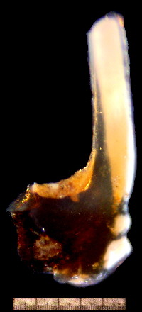
Transparent outer surface is actually striated with fine lamina, as demonstrated below on high-magnification images from the anterior edge of the tip, middle and base of the cusp. The lamina appear to maintain continuity along the length of the cusp, but outer layers are thinner at the base. Inner lamina merge with white matter at the tip -- which appears black in the high-power micrographs that use transmitted light. Wear surfaces buried inside the laminar layers suggest that the tooth actually grew continuously by adding new layers of enamel to the outer surface -- periodically recoating the worn element with a new and thicker layer, and simultaneously sharpening and repairing itself. Click on image to enlarge.
 -- BH1FBB13H04
Many very tiny inclusions appear in at the base of the white core of the cups.
 -- BH1FBB13H04.jpg